If you have been diagnosed with any of the below conditions and have an appointment with BCI soon, check out the ‘BEFORE YOUR FIRST APPOINTMENT’ page under the Patient Resources tab
Breast cancer is the most commonly diagnosed cancer amongst women in Australia. Approximately 57 Australians are diagnosed each and every day. That equates to over 20,000 Australians diagnosed with breast cancer each year.
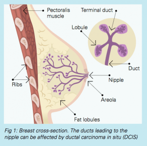
The normal breast
A woman’s breast is made up of 15 to 20 segments (lobes) in which there are smaller segments (lobules). These lobules are connected by tubes (ducts) that eventually join up and lead to main ducts at the nipple. The lobules are the milk producing parts of the breast. The ducts help to take the milk to the nipple during breastfeeding. In between the ducts and lobules there is fat and fibrous tissue holding all the structures together.
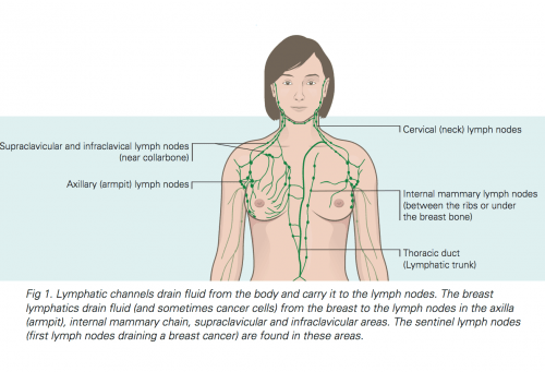
Lymphatic vessels are similar to veins, but carry lymph instead of blood. Lymph is a clear fluid that contains tissue waste products and immune system cells. Most lymphatic vessels of the breast lead to axillary (underarm) lymph nodes. Cancer cells may enter lymph vessels and spread out along these vessels to reach lymph nodes.
Lymph nodes are small, bean-shaped collections of immune system cells important in fighting infections. In some cases, breast cancer cells may reach the axillary lymph nodes via the lymph vessels and they can continue to grow, which may cause swelling of the lymph nodes in the armpit. If breast cancer cells have multiplied in the axillary lymph nodes, there is a chance that they may have spread to other parts of the body.
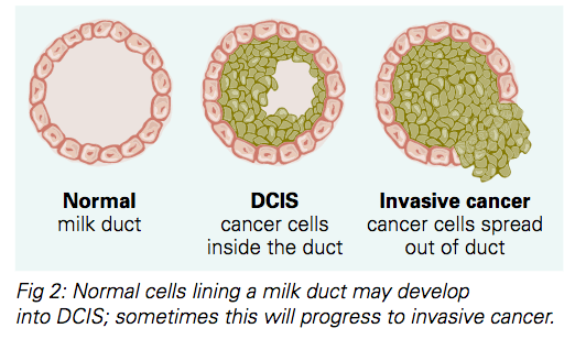
Carcinoma in Situ
Carcinoma in situ means that the cancer cells are within the duct or lobule and have not invaded the surrounding breast tissue.
There are two types of breast carcinoma in situ:
- Ductal carcinoma in situ (DCIS): The most common type of in situ breast cancer. The cancer cells grow inside the ducts and have not spread beyond it. Left untreated, DCIS will eventually spread into the surrounding tissue (invasive cancer).
- Lobular carcinoma in situ (LCIS): The condition is most often discovered as a result of a biopsy done for another reason, such as a suspicious breast lump or an abnormal mammogram. Similar to DCIS, if left untreated this may lead to an invasive cancer. Women with LCIS have an increased risk of developing invasive breast cancer in either breast, which may be duetal or lobular in type.
Invasive Carcinoma
Most invasive carcinomas are ductal carcinomas, which account for about 80% of all invasive carcinomas. Most of the remainder are invasive lobular carcinomas.
A diagnosis of cancer is usually given as a result of a biopsy (core biopsy or fine needle biopsy). This biopsy is examined by a pathologist under the microscope to confirm the diagnosis.
Many patients will be treated with surgery. The tissue removed at the time of surgery will be examined in detail by a pathologist.
The pathology report will contain detailed information on the type of the cancer, including the cancer grade (grade 1, grade 2 or grade 3), whether there is evidence of lymph node involvement, the surgical margins and whether hormone or HER2 receptors tests are positive. This will help the team of specialists plan specific treatment for each patient.